Case 1: Inferior Ischemia
Clinical History
Patient: 65-year-old male Presentation: Recurrent exertional chest pain Risk Factors: Hypertension, Hyperlipidemia, Former smoker Referral: Assessment of ischemia
Imaging Findings
Technique: One-day Rest/Stress Tc-99m Sestamibi SPECT/CT
Perfusion Images
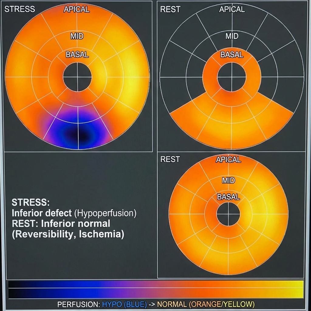
The myocardial perfusion polar map (bullseye plot) demonstrates:
- Stress: Moderate intensity defect involving the inferior and inferolateral walls (basal and mid segments).
- Rest: Significant improvement/normalization of the perfusion in the corresponding segments.
- Gating: Normal LV cavity size with normal wall motion and thickening. Calculated LVEF = 62%.
Diagnosis
Key Findings:
- Reversible perfusion defect in the inferior/inferolateral wall.
- Preserved wall motion and thickening.
Interpretation: Indicates ischemia in the distribution of the Right Coronary Artery (RCA).
Learning Points
- Reversibility: The hallmark of ischemia is a defect that is present at stress but improves or resolves at rest.
- Key Vascular Territories:
- LAD: Anterior wall, septum, apex
- RCA: Inferior wall, basal septum
- LCx: Lateral wall
- Prognostic Value: The extent and severity of ischemia (Summed Difference Score) correlates with future cardiac event risk.